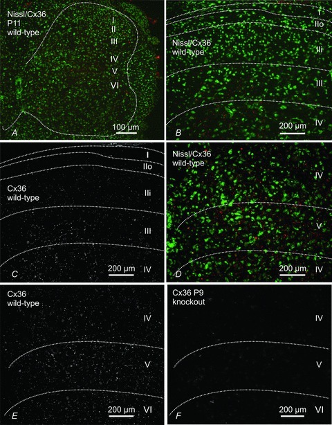
Figure 6. Immunofluorescence labelling for Cx36 in transverse sections of mouse L4 spinal cord in juvenile mice.

Panels A–E are from P11 mice and panel F is from a P9 mouse at the fourth lumbar segment. A, low magnification, fluorescence Nissl counterstained section (green) showing distribution of Cx36-positive puncta (red) in dorsal and ventral grey matter (outlined by dotted line). B and C, magnification of superficial dorsal horn laminae (outlined by dotted lines) with (B) and without (C) Nissl counterstain, showing sparse labelling for Cx36 (red) in lamina I and in outer (IIo) and inner (IIi) lamina II, and moderate labelling in lamina III. D and E, magnification of deeper dorsal horn laminae (outlined by dotted lines) with (D) and without (E) Nissl counterstain, showing abundant Cx36-puncta in laminae IV, V and a portion of VI. F, section from the dorsal horn of a P9 Cx36 knockout mouse showing absence of labelling for Cx36 in a field similar to that shown in E.
