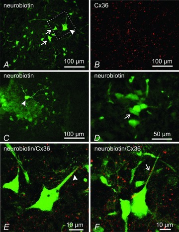
Figure 9. Immunofluorescence labelling of Cx36 (red) associated with neurobiotin-coupled neurones (green) in deep dorsal horn and intermediate zone of mice at P11.

A and B, low magnification showing a neurobiotin-injected neuron (A, arrowhead), giving rise to surrounding neurobiotin-positive neurons (A, arrows), and the same field (B) showing dense immunolabelling for Cx36 in lamina VI. C and D, neurobiotin-injected neuron in lamina V (C, arrowhead), and higher magnification from an adjacent section showing clusters of neurobiotin-positive neurones (D, arrow). E and F, confocal laser scanning images showing overlays of neurobiotin-positive neurones and immunofluorescence labelling for Cx36, with green/red overlap seen as yellow. The image in E is a magnification of the boxed area shown in A. Cx36-puncta are seen associated with dendrites and soma of a neurobiotin-injected neurone (E, arrowhead) and neurobiotin-coupled neurone (F, arrows).
