
Figure 6. KCNQ2 channels localize to nodes of Ranvier in mouse vagus nerve.
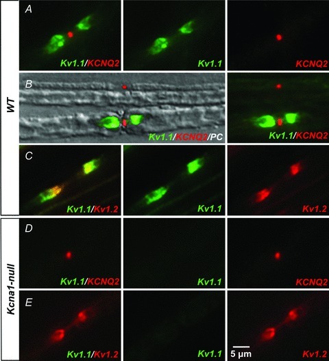
A, we detected KCNQ2 channels at nodes of myelinated vagal axons of 5-week-old wild-type mice using immunofluorescence. Co-labelling with anti-Kv1.1 antibodies showed Kv1.1-immunoreactivity at juxtaparanodes flanking the Kcnq2-positive nodes. B, Kv1.1-immunoreactivity appeared either weak or absent at many KCNQ2-positive nodes belonging to smaller fibres (<5 μm), as demonstrated in this phase contrast (PC) image superimposing Kv1.1- and KCNQ2-immunofluorescence. The small diameter (∼2 μm) fibre at the top shows only KCNQ2-positive nodal labelling, while the large diameter (∼7 μm) fibre at the bottom shows both Kv1.1-positive juxtaparanodal and KCNQ2-positive nodal immunoreactivity. C, Kv1.1-positive juxtaparanodes always exhibited Kv1.2-immunoreactivity. D and E, Kcna1-null fibres lack Kv1.1 labelling, but KCNQ2- and Kv1.2-immunoreactivity is preserved.