
Figure 4.
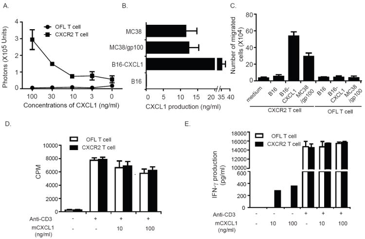
Enhanced migration of CXCR2-expressing T cells along CXCL1 gradients. (A) Migration of CXCR2-expressing T cells along recombinant CXCL1 gradients. C57BL/6 T cells were transduced with either OFL-GFP or OFL-GFP and mCXCR2 and placed into the upper wells of a Transwell system, with CXCL1 in the lower wells at the indicated concentrations. One hour later, D-luciferin was added to the lower wells. T cell migration to the lower well was quantitated by measuring luminescent activity. (B) CXCL1 production by murine tumor lines. Cells (2 × 10⁶) from the indicated murine cell lines were plated in 6-well plates; 24 h later, CXCL1 in supernatants was detected by ELISA. (C) Migration of CXCR2-expressing T cells along tumor cell-derived CXCL1 gradients. C57BL/6 T cells were transduced with either OFL-GFP or OFL-GFP and mCXCR2 and placed into the upper wells of a Transwell system. Conditioned cell culture medium from tumor cells was loaded into the lower wells. One hour after incubation, the cell number in the lower wells was counted. (D) Proliferation of CXCR2-expressing pmel-1 T cells upon TCR stimulation in the presence or absence of CXCL1. CXCR2-expressing T cells or OFL-expressing T cells were stimulated with anti-CD3 and irradiated splenocytes in the presence or absence of recombinant CXCL1. T-cell proliferation was evaluated by 3[H] thymidine incorporation assay. (E) IFN-γ secretion by CXCR2-expressing pmel-1 T cells upon TCR stimulation in the presence or absence of CXCL1. CXCR2-expressing T cells or OFL-expressing T cells were stimulated with anti-CD3 and irradiated splenocytes in the presence or absence of recombinant CXCL1.