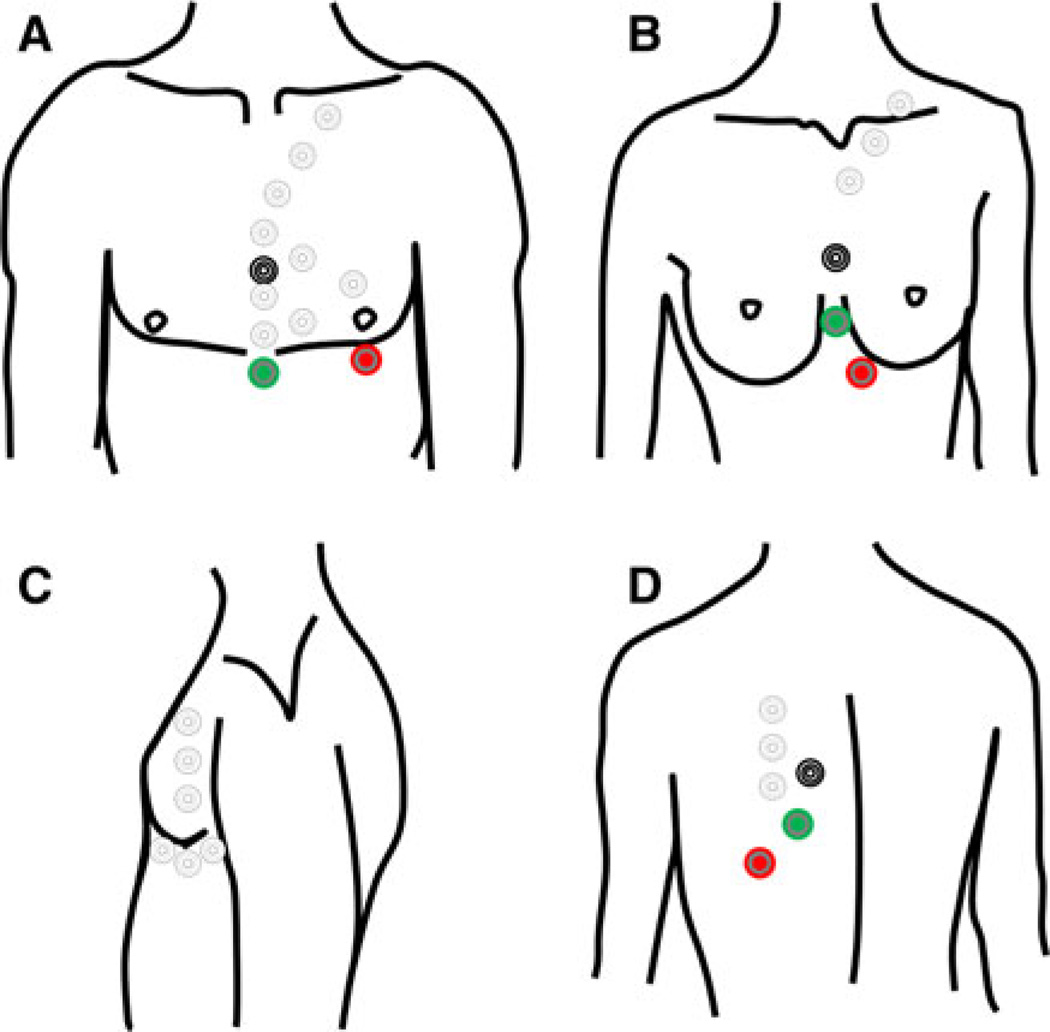
Fig. 1.
Black, green and red leads denote the standard position of the conventional electrodes. Uncolored circles represent alternative placements which can range from the mid-sternal region to the neck, lateral chest wall and back. a Frontal view. Alignment of the leads along the parasternal midline often increases the R-wave amplitude. b Frontal view. In women, it is often best to avoid the fatty breast tissue, so lead placement can be adapted to breast size and position. c Lateral positions. d Posterior view

