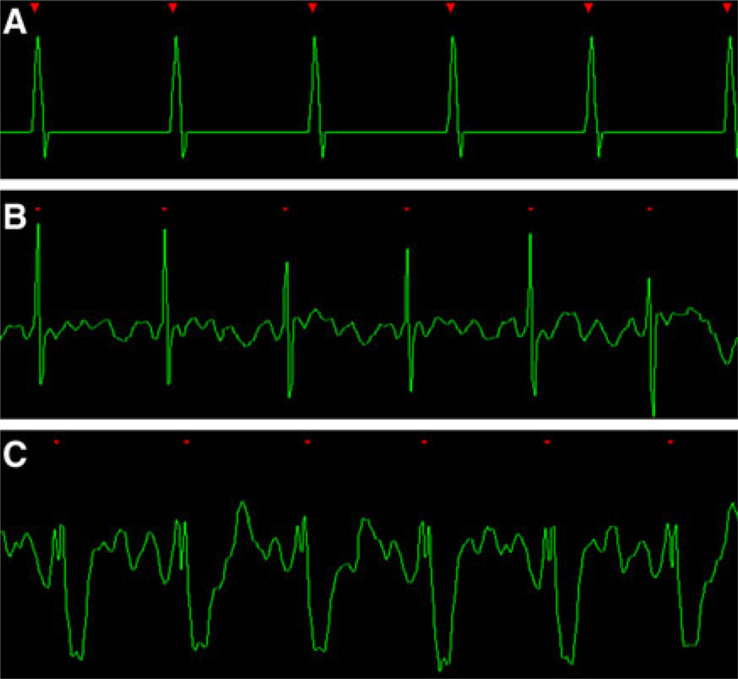
Fig. 2.
a An ideal ECG created by a simulation program. The R waves are tall and sensed every time, the R–R intervals are exactly evenly spaced and the intervening baseline is flat without any noise. b A excellent in vivo example. High peak amplitude R-waves are separated by minimal baseline noise. c The QRS complex is inverted; however, the high amplitude of the R-wave will serve as a reliable trigger for the acquisition

