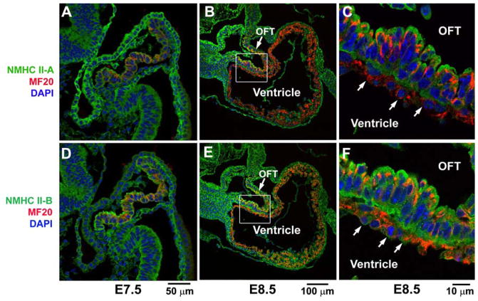
Figure 2. Expression of NM II during Early Heart Formation.
Immunofluorescence confocal images of wild-type mouse hearts at E7.5 and E8.5 stained for NMHC II-A (green, A–C) and II-B (green, D–F), and co-stained for MF20 (red, a marker for cardiac myocytes). At E7.5, the early cardiac myocytes (A,D) show co-staining of NMHC II-A and MF20 (A) or NMHC II-B and MF20 (D) indicating that both NMHC II-A and II-B are expressed in cardiac myocytes at this stage. At E8.5, the cardiac myocytes in the developing outflow tract (OFT) still express both NMHC II-A (B, magnified in C) and II-B (E, magnified in F), however in the ventricular myocytes at E8.5 (arrows, C,F) only NMHC II-B (E,F), and not NMHC II-A (B,C) is detected. Modified from reference 20.

