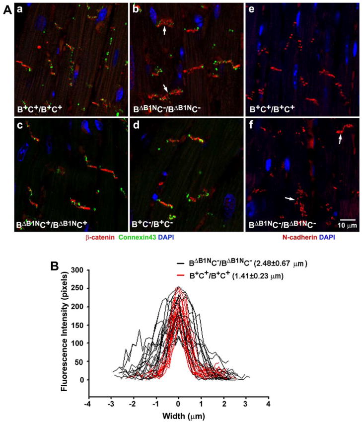
Figure 7. Impaired Localization of β-Catenin and N-cadherin in BΔB1NC−/BΔB1NC− Intercalated Discs.
(A) Immunofluorescence confocal images of adult heart sections from B+C+/B+C+, BΔB1NC−/BΔB1NC−, BΔB1NC+/BΔB1NC+, and B+C−/B+C− mice stained for β-catenin (red, a–d), connexin43 (green, a–d) and N-cadherin (red, e,f). In contrast to B+C+/B+C+ (a,e), B+C−/B+C− (d), and BΔB1NC+/BΔB1NC+ (c) cardiac myocytes where β-catenin and N-cadherin very precisely stain the intercalated discs, BΔB1NC−/BΔB1NC− myocytes (arrows, b,f) show a diffuse β-catenin and N-cadherin staining. No difference in connexin43 staining (green, a–d) is observed among these four genotypes. Nuclei were stained by DAPI (blue). (B) Profiles of β-catenin staining at the intercalated discs for B+C+/B+C+ (red lines) and BΔB1NC−/BΔB1NC− (black lines) mouse hearts. BΔB1NC−/BΔB1NC− hearts showed a diffuse β-catenin distribution manifested by widened β-catenin staining at the intercalated discs compared to the wild-type hearts. An embedded Zeiss LSM image profile tool is used to quantify the fluorescence intensity along a line drawn vertically across the intercalated discs. Reproduced from reference 20.

