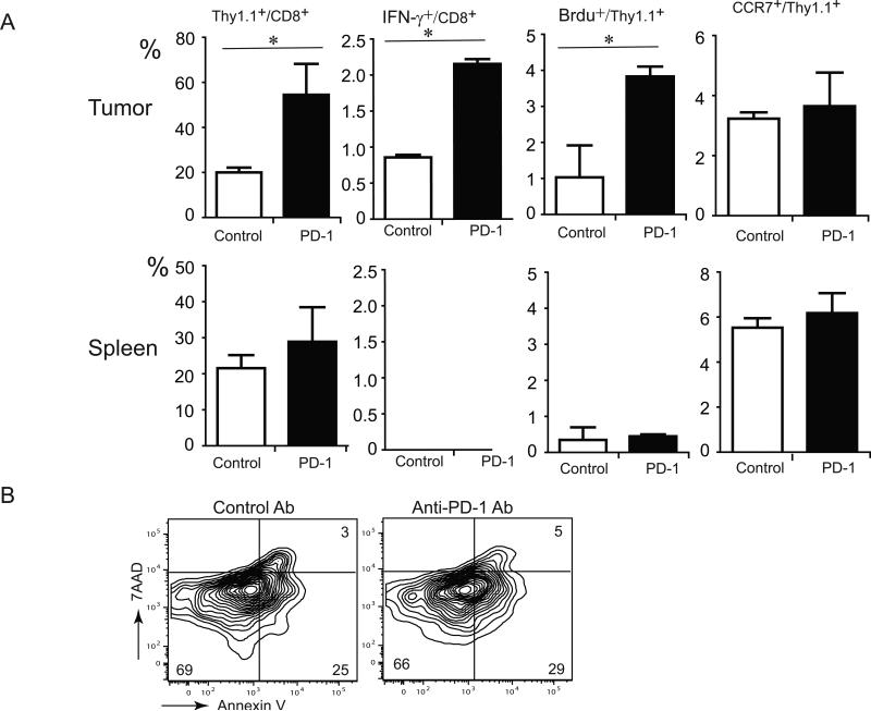
Figure 3.
The phenotype and function of transferred pmel-1 T cells in mice receiving ACT and anti-PD-1 Ab treatment. (A) Change of frequency and function of transferred pmel-1 T cells within tumor and spleen in response to PD-1 blockade. Six-days after T cell transfer, B16-bearing mice were intraperitoneally treated with Brdu solution. 24-hours later, mice were sacrificed to harvest tumor tissue and spleen (N=3 per group). Single cell suspension was made from tumor and spleen and stained with anti-CD8, anti-Thy1.1, anti-CCR7 anti-IFN-γ, and anti-Brdu. (B) Apoptosis of transferred pmel-1 T cells at the tumor site. Six-days after T cell transfer, lymphocytes from tumor tissues were stained with anti-CD8, anti-Thy1.1, Annexin V and 7AAD. Representative contour plots for one tissue sample from mice treated with ACT and anti-PD-1, as well as one tissue sample from mice treated with ACT with control antibody, were shown after gating with anti-Thy1.1+ and anti-CD8+ subsets. Values in each quadrant indicate the percentage of cells in the corresponding quadrant. (* indicates P<0.05)

