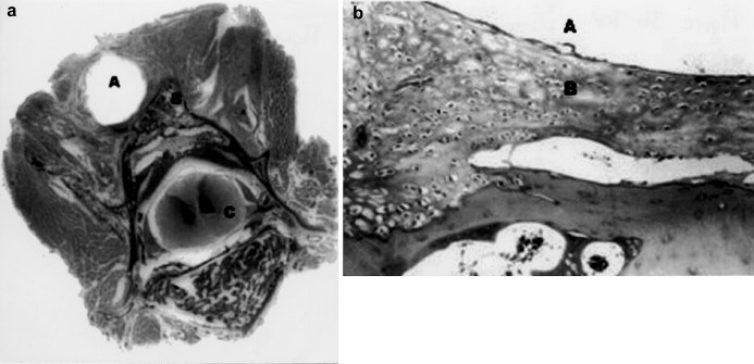
Fig. 3 a, b.
Sections of the paravertebral structures and vertebral body from an implanted animal from group 1 (a gross section original magnification; b hematoxylin-eosin stain, original magnification 40× ). A indicates the rod space; B indicates the fusion mass and C indicates the medulla spinalis

