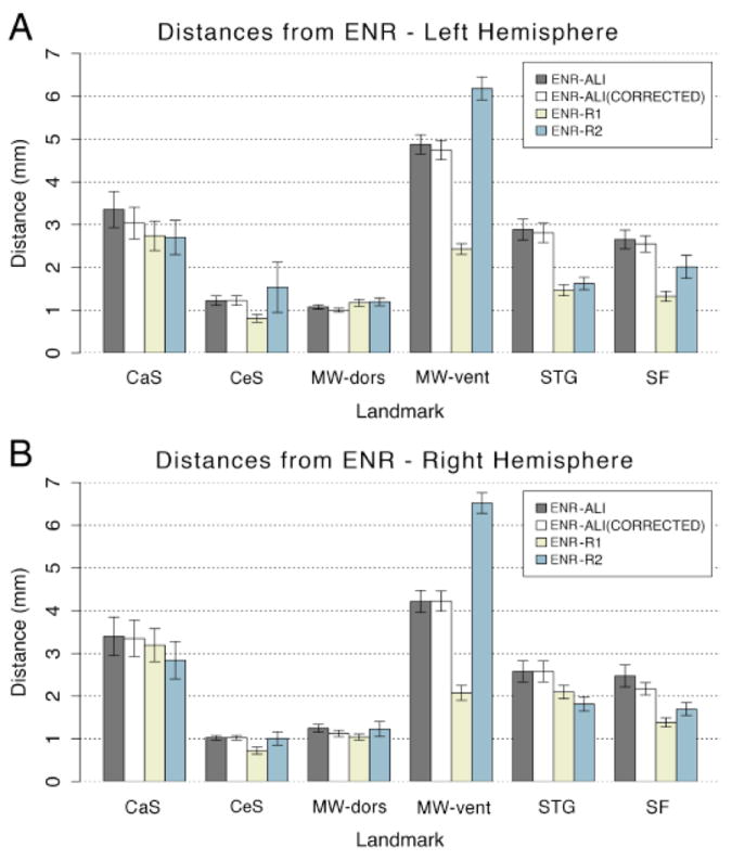
Figure 3.

Distances in mm from the expert neuroanatomical rater (ENR) are shown across all raters (ALI – automated landmark identification, R1 – Rater 1, R2 – Rater 2) and all landmarks (central suclus – CeS, calcarine sulcus - CaS, dorsal component of the medial wall – MW-dors, ventral component of the medial wall – MW-vent, superior temporal gyrus - STG, and sylvian fissure – SF). Results are shown for both (A) left and (B) right hemispheres. All distances were computed prior to SBR to avoid potentially obscuring differences post registration to the PALS-B12 atlas.
