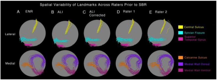
Figure 4.

Spatial variability of ‘Core 6’ landmarks prior to SBR is shown on a spherical configuration for the (A) expert neuroanatomical rater (ENR), (B) automated landmark identification (ALI), (C) ALI-Corrected, (D) Rater 1, and (E) Rater 2. The top panel shows the lateral view with central sulcus, sylvian fissure and superior temporal gyrus displayed in yellow, cyan and pink colors respectively. The bottom panel shows the medial view with calcarine sulcus, medial wall dorsal segment and medial wall ventral segment displayed in orange, dark and light purple respectively. The ‘clouds’ of variability allow for qualitative inspection of similarity across raters prior to surface registration to the PALS-B12 atlas.
