
Figure 1.
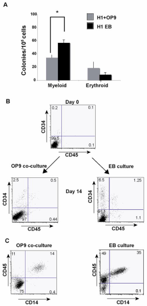
Embryoid body cultures yield more myeloid colonies as compared to OP9 cocultures : A) H1 cells co-cultured with OP9 feeders or as EB’s (in BASAL medium as shown in Table1) were dissociated into single cells following 14 days of culture. The single cells were plated in complete methylcellulose medium and analyzed for colony formation 2 weeks post plating. The figure is representative of the average of 3 independent experiments with OP9 feeders and 3 independent experiments as embryoid body cultures. Values of colonies that are significantly different between OP9 cultures and EB cultures are indicated by asterisks (*, P <0.0015). Probability values were calculated using Students t- test. (B) H1 cells co-cultured on OP9 feeders or as EB’s (in BASAL medium, Table1) were analyzed for expression of CD34 and CD14 at day 0 of culture. Following 14 days of culture in both conditions, cells were dissociated and analyzed for CD34 and CD45 expression by flow cytometry. (C) The CD34+ cells were sorted and differentiated further in presence of cytokines IL3, SCF and M-CSF and assayed for expression of CD14 and CD45. The figure is representative of 3 independent experiments.