
Figure 5.
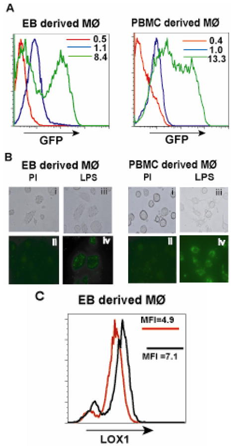
TLR ligands specifically increase phagocytosis of bacteria in EB derived macrophages similar to PBMC derived macrophages. (A) EB - derived Mφ’s and PBMC - derived Mφ’s were stimulated in the presence of LPS (green histograms) and in the absence of LPS (blue histograms) with media alone for 18hours. Following stimulation, the cells were challenged with E. coli expressing EGFP for 1.5 hrs and phagocytosis assayed by flow cytometry. Polyinosinic acid at a concentration of 0.5mg/ml was used as a negative control to inhibit phagocytic activity (red histograms). The MFI values are denoted in the histogram panels. (B), (C) The cells challenged with bacteria were analyzed by fluorescence microscopy. Panels (B) (i, ii) represent EB - derived Mφ’s treated with PI and (C) (i, ii) show PBMC - derived Mφ’s treated with PI. EB - derived Mφ’s treated with LPS are shown in panels (B) (iii, iv) while PBMC - derived Mφ’s are in panels (C) (iii, iv). Upper panels represent phase contrast images while lower panels represent fluorescent images. All images are at 200X magnification. (D) Upregulation of expression of LOX1 was analyzed by flow cytometry following treatment of cells with and without LPS. Data are representative of 2 independent experiments. Red histograms represent absence of LPS stimulation while black histograms represent cells treated with LPS. MFI values are shown within the histogram panels.