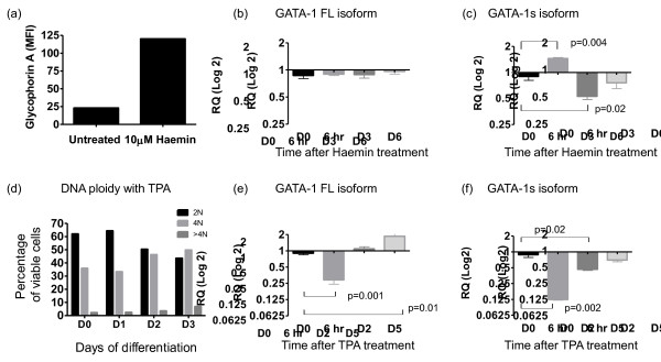
Figure 2.
GATA1s expression levels during haematopoietic differentiation of K562 cell lines by Haemin and TPA. (a) K562 surface glycophorin A expression measured by single colour FACS after 3 days of Haemin treatment (MFI = −Mean Fluorescence Intensity) (b, c) quantitative PCR analysis of (b) GATA1FL and (c) GATA1s, expression levels during the course of Haemin-induced erythroid differentiation (n = 3), results are expressed as fold change (log2) using day 0 as the calibrator (expression arbitrarily set at 1.0) (d) DNA ploidy analysis in K562 cells 0, 1, 2 and 3 days following treatment with the differentiating agent TPA (e, f) quantitative PCR analysis of (e) GATA1FL and (f) GATA1s, expression levels during the course of TPA-induced megakaryocytic differentiation (n = 3), results are expressed as fold change (log2) using day 0 as the calibrator (expression arbitrarily set at 1.0).

