Figure 1.
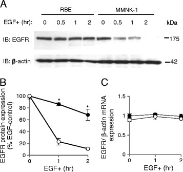
Epidermal growth factor receptor (EGFR) degradation upon EGF stimulation in RBE and MMNK-1 cells. (A) EGFR expression before and after 0.5, 1, and 2 hr of EGF treatment as detected by Western blotting. (B) Quantification of EGFR expression after 1 and 2 hr of EGF stimulation in RBE cells (closed circles) and MMNK-1 cells (open circles) from Western blotting. Values are standardized to the optical intensity of β-actin and presented as the percentage value of the EGFR expression value of RBE or MMNK-1 cells without EGF stimulation. *p < 0.05. (C) EGFR mRNA expression in RBE cells (closed circles) and MMNK-1 cells (open circles) before and after 1 and 2 hr of EGF stimulation as analyzed by RT-PCR. Values are standardized to β-actin mRNA expression and presented as the relative value to EGFR mRNA expression of RBE cells without EGF stimulation. All results shown are representative of four independent experiments.

