Figure 3.
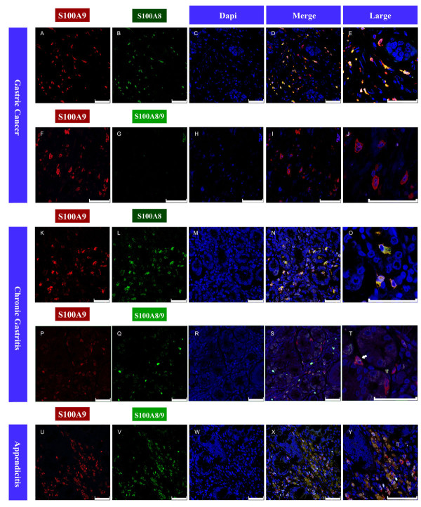
Immunofluorescence images of S100A9, S100A8 and S100A8/A9 proteins in tissue microarray slides containing gastric cancer tissues (A-J) and chronic gastritis tissues (K-T), and chronic appendicitis tissues with exacerbation (U-Y). S100A9 and S100A8 were detected by monoclonal antibody, prelabeled with the Zenon Alexa Fluor Mouse IgG Labeling Kit (with green and red fluorescence respectively). The nucleus was stained by DAPI. S100A8/A9 heterodimers were detectable using the dimer-specific antibody 27E10 from BMA Biomedicals prelabeled with green fluorescence. The co-localization of S100A9 and S100A8 or S100A8/A9 was showed in merged pictures (D, I, N, S, X) and larger merged pictures (E, J, O, T, Y). White arrow in 3 T shows co-localization of S100A9 and S100A8/A9 in chronic gastritis. Bar length, 50 μm.

