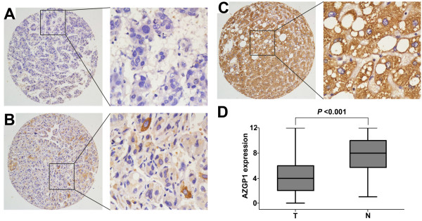
Figure 2.
The protein expression of AZGP1 in HCC by immunohistochemistry. The immunoreactivity was primarily observed in the cytoplasm within tumor cells. A. Negative staining of AZGP1 was detected in HCC case (#136) B. HCC case (#57) showed weak expression of AZGP1. C. Strong staining of AZGP1 was observed in 95% of the adjacent non-tumorous liver tissues. D. The box plot showed the mean staining score of AZGP1 in HCC tissues (T) and the adjacent non-tumorous liver tissues (N) (P < 0.001, t = -6.502).

