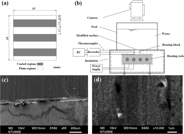
Figure 1.
Heating surface, experimental facility setup, and scanning electron microscopy (SEM) images. (a) Heating surface with coated and uncoated (plain) areas. (b) Experimental facility setup of the thermal system. (c) SEM image of nanoparticle-coated (downside, CA = 55°) and uncoated (plain) areas (upside, CA = 105°) at a scale bar of 200 μm. (d) SEM image of nanoparticles coated areas at a scale bar of, 1 μm.

