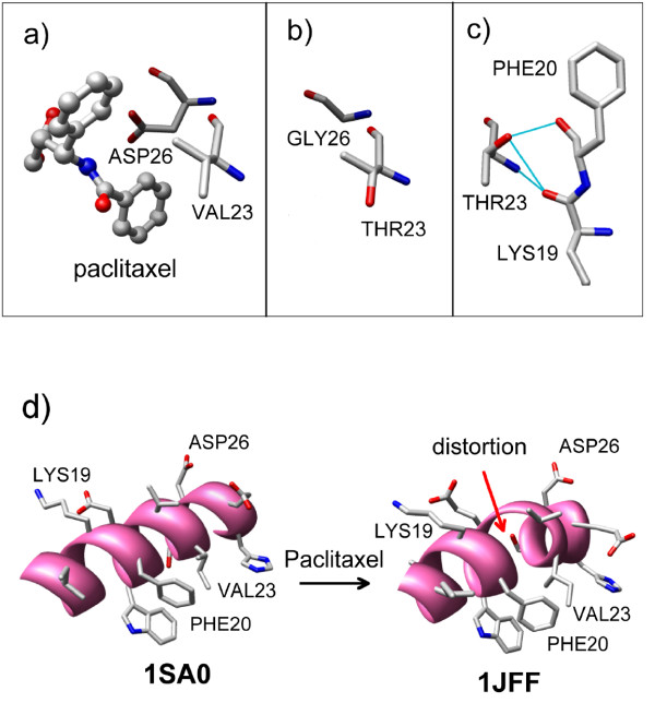
Figure 4.
Paclitaxel-bound (pdb ID:1JFF) and paclitaxel-free (pdb ID: 1SA0) β-tubulin. a) Disposition of Val23, Asp26 and paclitaxel in paclitaxel-bound β-tubulin (pdb ID:1JFF). b) Same as panel (a) with two in silico mutations: Val23Thr and Asp26Gly. The rotameric state of Val23 in the original pdb file (1JFF) was maintained during the mutation. c) Identical to panel (b) except that the side-chain of Thr23 has been changed to a new rotameric state. In the new rotameric state, the sidechain of Thr23 can form a H-bond with the backbone carbonyl oxygen atoms of Phe20 and Lys19. d) An α-helix in β-tubulin (containing residues 19, 20 and 23) as found in two crystal structures: 1SA0 (paclitaxel-free) and 1JFF (paclitaxel-bound).

