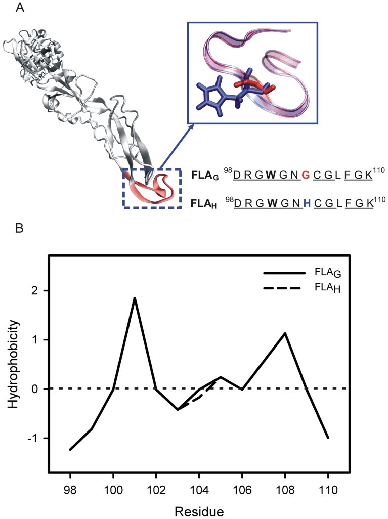
Figure 1. The flavivirus E glycoprotein fusion loop and hydrophobicity plots of the fusion peptides.
(A) The crystallographic structure of West Nile virus E protein (PDB ID 2HG0) and the schematic representation and sequence of the two fusion peptides of flaviviruses studied in this work. FLAG has a Gly residue (red) in position 104 of glycoprotein E, while FLAH presents a His residue (blue). Trp101, Gly104 and His104 are indicated in bold, red and blue, respectively. The conserved amino acids are underlined. (B) Hydrophobicity plots for the fusion peptides FLAG (solid line) and FLAH (dashed line) were elaborated using the Wimley-White hydrophobicity scale.

