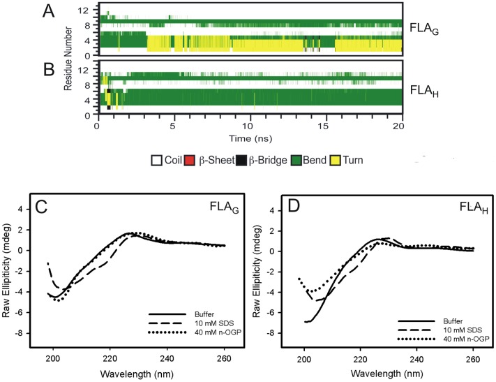
Figure 6. The secondary structures of FLAG and FLAH in the presence of membrane models.
(A and B) The secondary structures of the fusion peptides FLAG (A) and FLAH (B) in the presence of POPE membranes at 35°C. (C and D) The circular dichroism spectra of the FLAG (C) and FLAH (D) FP in solution (solid line) and in the presence of SDS (dashed line) or n-OGP (dotted line) micelles. The experiments were performed at room temperature at pH 5.5.

