
Figure 7. Increased neutrophils in spleen of Npc1 −/− mice. (A).
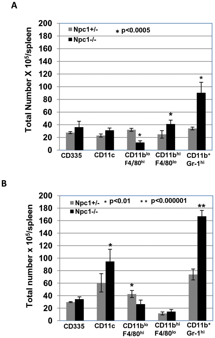
Flow cytometric analysis of innate immune cells in spleen of Npc1+/− and Npc1−/− mice. Splenocytes from un-infected Npc1+/− and Npc1−/− female littermates (age 6–8 weeks) mice were isolated and stained with anti-CD335 for NK cells, anti-CD11c for dendritic cells (DC), anti-F4/80 and CD11b for monocytes and macrophages (Mo/MO), anti-Gr-1 and CD11b for neutrophils. The data represent the mean from two independent experiments with a total of 6 mice (3 each experiment). Error bars show the mean±SD. Gating parameters are indicated in Figure S3. (B) Flow cytometric analysis of innate immune cells in the spleen of S. typhimurium infected Npc1+/− and Npc1−/− mice. Mice at 6–8 weeks were infected with S. typhimurium intraperitoneally (see Materials and Methods) and splenocytes were prepared at 48 hpi. Innate immune cells were analyzed as described in A. The data represent the mean from three independent experiments with a total of 6 mice (2 each experiment). Error bars indicate the mean±SD. Statistical significance was determined using Student’s t test. Gating parameters are indicated in Figure S3.