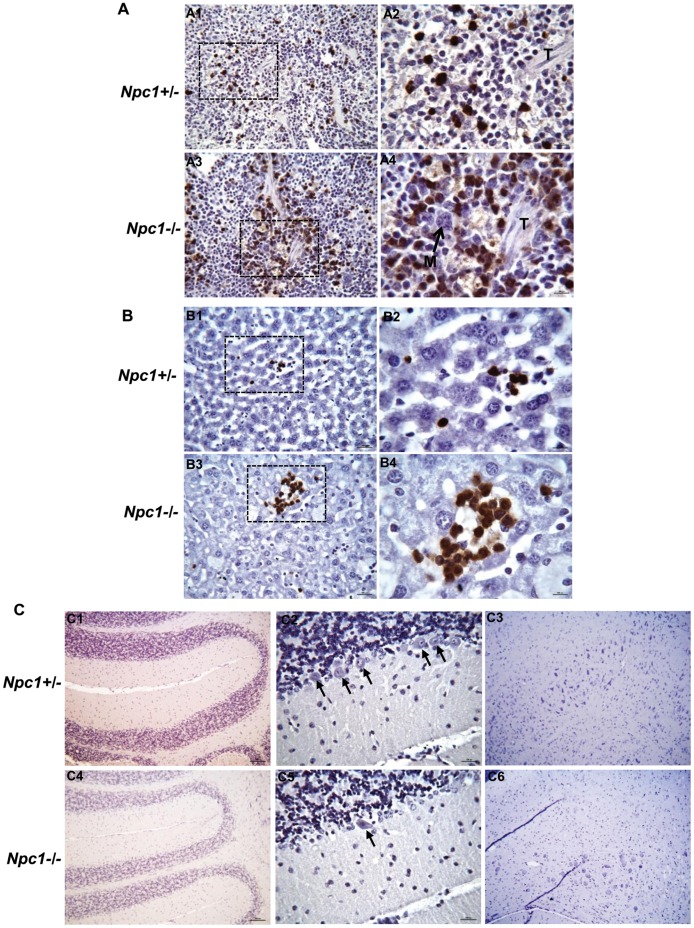
Figure 8. Immunohistochemical analyses of spleen, liver and brain sections.
(A) Immunohistochemical analyses reveal increased accumulation of neutrophils in the spleen of Npc1−/− mouse. Formalin-fixed paraffin embedded spleen sections (3–4 µm) of Npc1−/− and Npc1+/− mice (age 48–52 days) were stained with anti-Gr-1 antibodies to visualize neutrophils (cells stained in brown) which were primarily observed in the marginal zone, and in the red pulp of the spleen. Prominent accumulation of neutrophils was seen in the red pulp of Npc1−/− mouse (A3–4) compared to Npc1+/− mouse (A1–2). A2 and A4 are magnified view of area shown by dotted box in A1 and A3 respectively. M, megacaryocyte; T, trabecula. Original magnifications, x400 (A1&A3) and x1000 (A2&A4). (B) Detection of giant foci of neutrophils (cells stained in brown) in the liver of Npc1−/− mouse age 48–52 days (B3–4). These large foci of neutrophils were not detected in the liver of age-matched Npc1 +/− mouse (B1–2). Immunohistochemical staining on formalin-fixed paraffin embedded liver sections (3–4 µm) were carried out using anti-Gr-1 antibodies to visualize neutrophils. Tissue damage is clearly evident (B3–4) in the area of neutrophils accumulation in Npc1−/− mouse. B2 and B4 are magnified views of areas shown by dotted boxes in B1 and B3 respectively. Original magnifications, x400 (B1&B3) and x1000 (B2&B4). (C) Immunohistochemical staining of formalin-fixed paraffin embedded brain sections of Npc1 −/− and Npc1 +/− mice (age 48–52 days) was performed using anti-Gr-1antibodies. The entire brain (sagittal sections) was scanned. Panels are; C1and C4, cerebellum of Npc1+/− and Npc1 −/− mouse respectively; C2 and C5, magnified view of cerebellum of Npc1+/− and Npc1 −/− mouse respectively; C3 and C6, magnified view of regions from mid brain of Npc1+/− and Npc1 −/− mouse respectively. Several purkinje cells are evident (shown by black arrows) in Npc1 +/− (C2), however in Npc1−/− (C5) only few are seen. Original magnifications, x100 (C1, C3, C4 & C6) and x400 (C2&C5).

