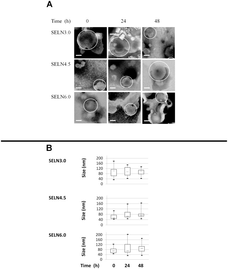
Figure 1. Synthetic exosome-like nanoparticles (SELN).
(A) Synthetic exosome-like nanoparticles (SELN) were examined by electron microscopy. SELN3.0, SELN4.5 and SELN6.0 preparations were incubated at room temperature, then 3 µl were removed at 0, 24 h and 48 h. SELN were then disposed on top of Formvar-coated 300-mesh carbon grids and treated with 2% phosphotungstic acid. Several fields were photographed and used to determine the diameter of SELN. Scale bars on microphotographs represent 50 nm. (B) The range of observed diameters for lipid structures as represented in A was statistically represented by the box plot. The dashed line inside the box represents the median diameter of SELN (n = 20). The box represents the interquartile range (50% of values). Tails extend to values within 1.5 times the interquartile range.

