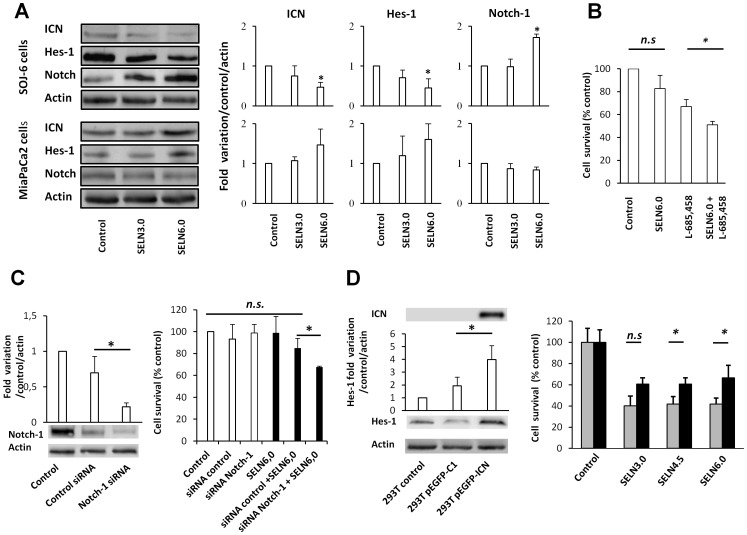
Figure 3. Effect of SELN on Notch pathway.
(A) SOJ-6 and MiaPaCa-2 cells were starved then treated for 24 h with SELN (16 nmoles cholesterol/ml) and lysed. Cell lysate proteins were separated on SDS-PAGE (80 µg of proteins/lane) and electrotransferred onto nitrocellulose membrane. The levels of ICN, Hes-1, Notch-1 and β-actin were determined by probing membranes with specific antibodies as indicated. Western blots are representative of three independent experiments. Lane 1, control performed in the absence of SELN, Lane 2, in the presence of SELN 3.0, Lane 3, in the presence of SELN 6.0. The right panels in figure 3A represents the quantification of western blots (means (± SD) of three independent experiments) using the NIH Image program (Mann-Whitney test). (B) MiaPaCa-2 cells were incubated with medium (control), with L-685,458 γ-secretase inhibitor (GSI, 2.5 µM), with SELN6.0 (16 nmoles of cholesterol/ml), with SELN6.0 and GSI. The L-685,458 GSI was added 60 min before freshly prepared SELN6.0. Cells were further incubated for 24 h in the presence of each component and cell proliferation was determined. Results are means (± SD) of independent experiments (n = 36, Student’s t-test). (C) MiaPaCa2 cells were transfected with a mix of Notch-1 siRNA or with a control siRNA. In the left panel cell lysate proteins (50 µg of proteins) of parental, control and Notch-1 siRNA transfected MiaPaCa-2 cells were separated on SDS-PAGE and analyzed by western blotting (72 h post-transfection). The histogram indicates quantification of western blots. In the right panel cell survival of parental and transfected MiaPaCa-2 cells was assessed through a MTT test after 24 hours of incubation with (black columns) or without (white columns) SELN6.0. The proliferation of MiaPaCa-2 cells recorded in the absence of SELN was taken as 100%. Results are means (± SD) of three independent experiments (Mann-Whitney test). (D, left panel) HEK 293 T cells were transiently transfected with the pEGFP-C1 control plasmid or pEGFP-ICN plasmid encoding ICN (Notch-1 intracellular domain). Cell lysate proteins of parental and transfected HEK 293T cells were separated on SDS-PAGE and analyzed by western blotting using primary antibodies as indicated. Western blotting replicates depicted the expression level of ICN and the nuclear target of ICN, Hes-1, in parental (293T control), pEGFP-C1 control vector-transfected (293TpEGFP-C1) and pEGFP-ICN-transfected (293TpEGFP-ICN) HEK 293T cells. The amount of loaded proteins was the same (50 µg) for each lane as indicated by β-actin probing. The histogram displays the quantification of Hes-1 as determined from western blots. (D, right panel) HEK 293TpEGFP-C1-transfected cells (grey columns) and HEK 293TpEGFP-ICN-transfected cells (black columns) were challenged without (control) or with SELN3.0, SELN4.5 and SELN6.0 (16 nmoles cholesterol/ml) for 24 h. Cell survival was determined with MTT. The proliferation of HEK 293T cells transfected with the control vector (293TpEGFP-C1) recorded in the absence of SELN was taken as 100%. Values are means ± SD of three independent experiments (Mann-Whitney test).

