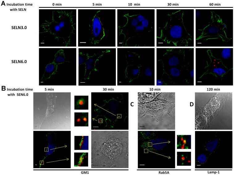
Figure 10. Fluorescent SELN incorporation.
(A) SOJ-6 cells were seeded on 1.4 cm-diameter cover slips in 12-wells plate, once adherent cells were starved for 24h before incubation with SELN3.0 or SELN6.0 (8 to 10 µl of SELN solution corresponding to 1.6 nmol cholesterol in 100 µl culture medium). SELN used in this experiment were only loaded with N- Rh-PE. Cells were incubated for 0, 5, 10, 30 and 60 min with N- Rh-PE-loaded SELN. At the end of the incubation time cells were washed with PBS and then fixed and saturated, nuclei were blue-colored with Draq5 (1 µM, 10 min, 37°C) and saturated with 1% BSA. Cell plasma membranes were further labelled with mAb16D10, a monoclonal antibody which binds to the tumor cell membrane antigen 16D10 [25]. mAb16D10 is then detected using a secondary antibody to IgM, coupled to FITC. Examination was performed using a SP5 Leica confocal microscope (Scale bar = 5 µm). (B) Plasma membrane lipid microdomains were visualized via the binding of the cholera toxin subunit B (CT-B) to raft ganglioside GM1. SOJ-6 cells were grown in complete DMEM medium, incubated with SELN6-Rh-PE (5 min, 37°C in FCS-depleted DMEM) before washing twice. Cells were then fixed with PFA, and nuclei were blue-colored with Draq5. Fixed cells were washed and incubated with the CT-B (0.5 µg/ml final concentration, 10 min, 4°C) before washed and incubated with Alexa Fluor 488–conjugated antibodies against CT-B (15 min, 4°C) (GM1, 5 min). To detect the intracellular localization of the CT-B, cells were first incubated with CT-B (see above), washed, and incubated with Alexa Fluor 488–conjugated antibodies against CT-B, before incubation with SELN6.0-Rh-PE, during 30 min at 37°C. Finally cells were fixed with PFA and then nuclei labelled with Draq5 (GM1, 30 min). (C, D) SOJ-6 cells were treated as in A and incubated (for the indicated time) with SELN6.0 labelled with N-Rh-PE (1.6 nmol cholesterol/100 µl culture medium). Cells were fixed with PFA and nuclei labelled with Draq5. Cells were permeabilized with saponin (0.1%, 30 min at room temperature), saturated (BSA, 1%, 30 min at room temperature) and incubated with antibodies (C) to early-endosome marker Rab5A, or (D) to late endosome marker Lamp-1 and further detected with an Alexa Fluor 488-labelled secondary antibodies. In B and C, squares indicate co-localization (yellow) and arrows indicate enlarged areas (inserts) where GM1, or Rab5A co-localizes with N- Rh-PE labelled SELN6.0 (Scale bar = 5 µm).

