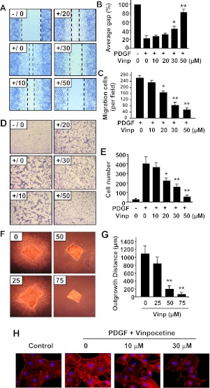
Fig. 4.
Effects of vinpocetine on VSMC migration. A to C, scratch wound assay. A, representative images show that vinpocetine dose-dependently inhibited PDGF-induced migration of VSMCs by scratch wound assay. Dotted lines show the edges of cell migration. Confluent VSMCs were starved and scratched with a pipette tip, then treated with vinpocetine at the indicated doses and stimulated with 25 ng/ml of PDGF-BB for 16 h. The cells were fixed with 4% paraformaldehyde and stained with hematoxylin. B and C, quantitative data of the scratch wound assay were analyzed by the percentage of gap area (B) or migrating cell numbers (C). D and E, Boyden chamber assay. D, representative images show that vinpocetine dose-dependently inhibited PDGF-induced transmigration of VSMCs by Boyden chamber assay. One hundred microliters of VSMC suspension was placed in the upper microchemotaxis chamber, and 600 μl of DMEM containing 25 ng/ml of PDGF-BB was placed in the lower polycarbonate filter chamber. The chamber was incubated at 37°C and 5% CO2 for 6 h. E, the transmigrated cells on the filter membrane were fixed and stained with hematoxylin and quantified. F and G, ex vivo aortic medial explant migration assay. F, representative images show that vinpocetine dose-dependently inhibited PDGF-induced VSMC outgrowth in 3D collagen I gel. Media explants of mouse aorta were embedded in 3D gel containing collagen type I, and the migration of VSMCs was initiated by addition of PDGF-BB/FGF2. G, migration was quantified by measuring the distance migrated by the leading front of VSMCs from the explanted tissue. Values are means ± S.D. from at least three independent experiments. *, P < 0.05; **, P < 0.01 versus PDGF with no vinpocetine. H, effects of vinpocetine on actin cytoskeleton polymerization in VSMCs. VSMCs were seeded in six-well plates overnight in DMEM supplemented with 10% FBS, serum-starved for 48 h, and treated with or without vinpocetine for 0.5 h, then stimulated with 20 ng/ml of PDGF-BB for 24 h. F-actin was stained by Alexa Fluor 546 phalloidin (red), and nucleus was stained by DAPI (blue).

