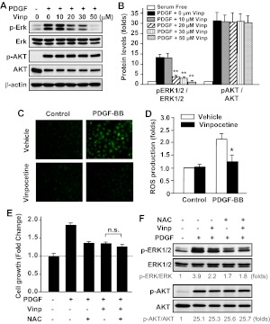
Fig. 6.
Effects of vinpocetine on PDGF-induced ERK1/2 and AKT phosphorylation and ROS production in VSMCs. A and B, VSMCs were serum-starved for 48 h, then treated with vinpocetine at the indicated doses and stimulated with 20 ng/ml of PDGF-BB for 15 min. ERK1/2 (A) and AKT (B) phosphorylation and total ERK1/2 and AKT levels were measured by Western blotting with phospho-specific antibodies and total antibodies, respectively. **, P < 0.01 versus PDGF with no vinpocetine. C and D, serum-starved VSMCs were labeled with 10 μM DCFH2-DA, then treated with vinpocetine and stimulated with 20 ng/ml of PDGF-BB for 1 h. C, the photographs were taken by using an Olympus (BX-51) fluorescent microscope. D, intracellular ROS was quantified by flow cytometry. *, P < 0.05 versus PDGF with vehicle. E, serum-starved VSMCs were pretreated with 30 μM vinpocetine, 5 mM NAC, or both, followed by PDGF-BB (50 ng/ml) stimulation for 48 h. Cell growth was measured by SRB assay. n.s., not significant. F, serum-starved VSMCs were pretreated with 30 μM vinpocetine, 5 mM NAC, or both, followed by PDGF-BB (50 ng/ml) stimulation for 15 min. ERK1/2 and AKT phosphorylation and total ERK1/2 and AKT levels were measured by Western blotting. The blots were analyzed by densitometry. Fold changes normalized to the left lane are shown below the blots (n = 2–3).

