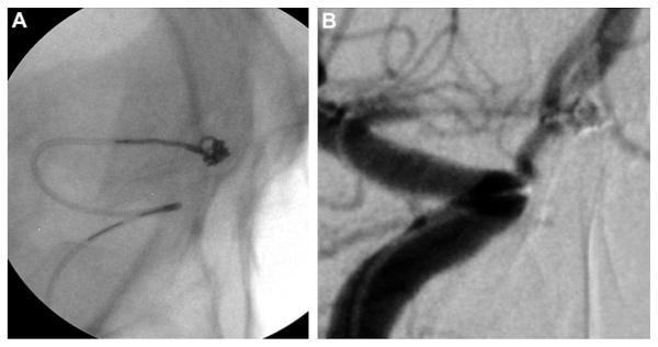
Figure 2.
(A) Still image during coil embolization with use of microangiographic fluoroscope detailing coil, the microcatheter and its distal marker, and the interface between the microcatheter and the coil. (B) Working anteroposterior view during coil embolization with standard fluoroscopy. Compared with (A), the resolution of the microcatheter and the coil is much lower.

