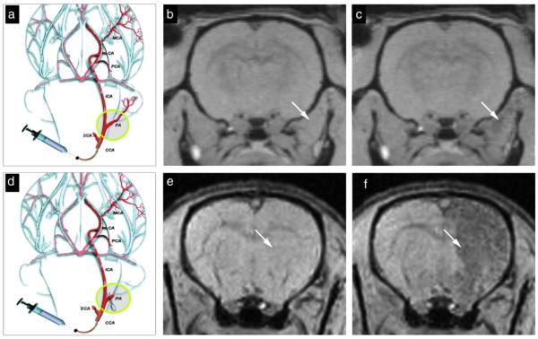
Fig. 1.
Real-time MR monitoring of injection accuracy. Following ligation of the external carotid and occipital arteries, the common carotid artery was cannulated (a) and SPIO-labeled cells were infused. In this experiment, the pterygopalatine artery was left intact. MR images were acquired immediately pre-injection (b) and post-injection (c). MR images demonstrate that the vast majority of cells localized into the extracerebral tissue, with negligible binding within the brain. When the pterygopalatine artery was ligated (d), all infused cells were perfused into the internal carotid artery and localized successfully into the ipsilateral hemisphere. Shown are the MR images acquired immediately before (e) and after injection (f). Reproduced, with permission, from Ref. [113].

