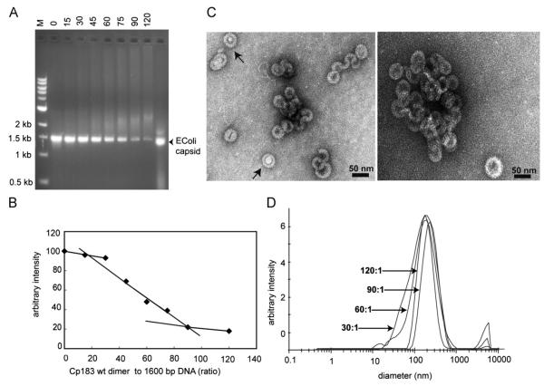
Fig. 4. Binding of 1600 bp dsDNA by Cp183 dimer.
(A) EMSA of 1600 bp dsDNA titrated with Cp183 dimer at the listed molar ratio of dimer to dsDNA polymer. (B) A plot of unbound 1600 bp dsDNA from EMSA, quantified with image J, showing its disappearance as a function of protein concentration. (C) Negative stain micrographs of Cp assembled on a 1600 bp dsDNA associated with core protein at ratios of 60:1 (left) and 120:1 (right); the scale bar is 50 nm. (D) Size distribution of Cp183-dsDNA complexes, determined by DLS, increases as the Cp183:dsDNA ratio increases from 30:1 to 120:1; the respective centroids of diameter distributions are 117 nm, 137 nm, 146 nm, and 155 nm.

