Abstract
A prospective study of 100 consecutive patients of deviated nasal septum to analyze association of septal deviation with external nasal deformity was undertaken at Acharya Vinoba Bhave rural Hospital of Jawaharlal Nehru Medical College, Sawangi (Meghe) Wardha from January 2009 to September 2010. Nasal septal deviations were evaluated by clinical examination and diagnostic nasal endoscopy while external nasal deformities, after evaluating, were documented using high resolution photography Nasal septal deviations were classified in seven types from I to VII by using Mladina’s classification modified by Janardhan et al. Jang classification was employed to classify external nasal deformities. 66% of the patients with deviated nasal septum were symptomatic while 34 lacked symptoms. Nasal obstruction was the most frequent symptom in 64% followed by nasal discharge in 33% Type VII was the most common type of deviation in 29%. Study revealed that 67% of the patients with deviated nasal septum had external nasal deformity and of the 67 patients with external deformity, Type I deformity was most frequent (26%). Remarkable feature of our study was Type I, III, V septal deviations were not associated with external deviation Type II septal deviations were commonly associated with Type III external deformity (7%) and Type IV septal deviation were closely associated with Type I external deformity (12%).
Keywords: Deviated nasal septum, External nasal deviation
Introduction
Pivotal role of nasal framework, a central structure of the face, in imparting beauty and pleasant look can not be overemphasized. Monumental statement made by Beckhius [1] ‘As goes the septum so goes the nose’ speaks profusely on the indispensable role of nasal septum in supporting the framework. Nasal septal deviations which quite often, if not always, implies external deformity, can adversely affect facial aesthetics and harmony. Thus a curved or deviated nasal septum become clinically significant when it results in infirmity either in form or function.
Whenever there is an external deformity of nose, its anatomical basis may be rooted in bony pyramid defect, cartilaginous framework defect, septal deformity or combination of these vectors. The crooked and scoliotic nose is a frequent reason for patient seeking aesthetic correction. On the part of rhinologists, these deformities, calls for extremely astute approach and acumen to diagnose and analyze both, the subtle and gross facets of the deformity.
It’s not only form but function which makes patient attend rhinology services.
Septal deviations with or without external deformities can lead to symptoms ranging from nasal obstruction to nasal bleed. Apart from vital role of deviated nasal septum in perpetuating sino nasal infections, it can also negatively contribute to common maladies like headache and allergy.
In the present study, attempt has been made to critically analyze 100 patients of deviated nasal septum. The main thrust of the study was on classifying nasal septal deviations and external deformities and correlating them.
Materials and Methods
A total of 100 patients between the age group of 16–65 years attending the ENT OPD of Acharya Vinoba Bhave Rural Hospital of Jawaharlal Nehru Medical college Sawangi (Meghe), Wardha, Maharashtra, India were accrued for this study. This prospective study was carried out between January 2009 to September 2010.
Inclusion criteria for the study were patients of rural background of Maharashtra presenting with external nasal deformity, deviated nasal septum, septal dislocation, septal spur, septal thickening and septal perforation external deformity due to other conditions like granulomatous diseases and malignancies were excluded. Also excluded were the patients with septal haematoma, abscess, sinonasal mass and subject belonging to urban area.
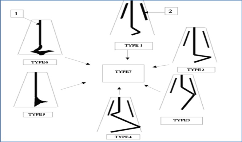
These patient with deviated nasal septum with or without external nasal deformity were evaluated clinically, using anterior rhinoscopy and diagnostic nasal endoscopy (0 and 30 degree) for morphology of nasal septum external deformity were documented by standard high resolution photographic analysis. Septal deformities were classified using Mladina classification modified by Janardhan et al. [2, 3] which classifies septal deformities into seven types. In this classification Type I–VI are separate entities while Type VII is combination of Type I–VI (Fig. 1; Table 1).
Fig. 1.
Mladina’s classification of nasal septal
Table 1.
Mladina’s classification of nasal septal deviation
| Type I | Mild deviation in vertical plane |
| Type II | Moderate anterior vertical deviation of cartilaginous septum in full length |
| Type III | Posterior vertical deviation at level of OM and middle turbinate |
| Type IV | ‘S’-shaped, posterior to one side and anterior to other |
| Type V | Horizontal septal crest touching or not touching the lateral nasal wall |
| Type VI | Prominent maxillary crest contralateral to the deviation with a septal crest on the deviated side |
| Type VII | Combination of previously described septal deformity types |
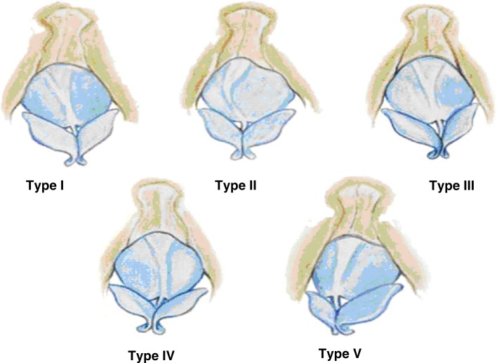
External nasal deformities were classified employing Yong Jo Jang’s classification [4]. According to this classification external nasal deformities are classified into five types based on the orientation of bony pyramid and cartilaginous vault to each other (Fig. 2; Table 2).
Fig. 2.
Yong Ju Jang’s classification for external nasal deformity (Type I–V)
Table 2.
Yong Ju Jang’s classification of external nasal deformity
| Type | Description |
|---|---|
| I | Straight tilted bony pyramid with tilted cartilaginous vault in the opposite direction |
| II | Straight tilted bony pyramid with concavely or convexly bent cartilaginous vault |
| III | Straight bony pyramid with tilted cartilaginous vault |
| IV | Straight bony pyramid with bent cartilaginous vault |
| V | Straight tilted bony pyramid and tilted cartilaginous dorsum in the same direction |
This data along with the demographic details and photographic documentation was collected and analyzed statistically using Chi-square test. The findings are being presented here.
Results
We looked at a total of 100 patients in the age group of 16–65. Mean age of the patient was 34.7 years. Maximum number of patients were in the age bracket of 31–40 (34%) followed by 29% in the age group of 21–30 (Table 3). 63 patients (63%) were males and 37 (37%) were females. Male to female ratio was thus 1.7:1.
Table 3.
Age wise distribution of patients
| Age group (years) | No. of patients |
|---|---|
| 10–20 | 9 |
| 21–30 | 29 |
| 31–40 | 34 |
| 41–50 | 21 |
| >50 | 7 |
| Total | 100 |
66 (66%) patients in the present study were symptomatic while the rest 34 (34%) lacked symptoms (Fig. 3). Nasal obstruction (64%) was the leading nasal symptom followed by nasal discharge (33%).
Fig. 3.
Distribution of patients according to symptoms
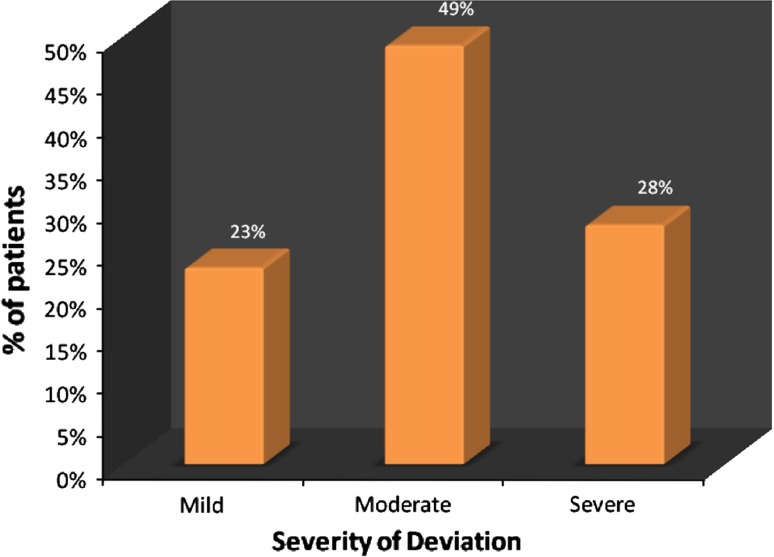
Morphologically septal deviations in our study were classified into seven types by adopting a classification evolved and described by Mladina [2] and modified by Janardhan et al. [3] (Fig. 1; Table 1). Incidence of Type VII was most common (29%) followed by Type IV (22%) (Table 4). On correlating the symptoms with the type of deviation it was found that Type VII septal deviation was most common (20%) among symptomatic group. This was followed by Type IV in 18%. In asymptomatic group Type II septal deviation was more common. Severity of deviation was classified into mild, moderate and severe based on distance of deviated septum from lateral nasal wall on the convex side. 49% patients had moderate deviation. This is given in Fig. 4.
Table 4.
Distribution of patients according to type of nasal septal deviation
| Type of nasal septal deviation | No. of patients | Percentage |
|---|---|---|
| I- Mild deviation in vertical plane not extending throughout entire length of septum | 5 | 5 |
| II- Moderate anterior vertical deviation of cartilaginous Septum | 21 | 21 |
| III- Posterior vertical deviation at level of OM and middle turbinate | 5 | 5 |
| IV-‘S’-shaped, posterior to one side and anterior to other | 22 | 22 |
| V- Horizontal septal crest touching or not touching the lateral nasal wall | 7 | 7 |
| VI- Prominent maxillary crest contralateral to the deviation with a septal crest on the deviated side | 11 | 11 |
| VII- Combination of described septal deformity types | 29 | 29 |
| Total | 100 | 100 |
Fig. 4.
Distribution of patients according to severity of deviation
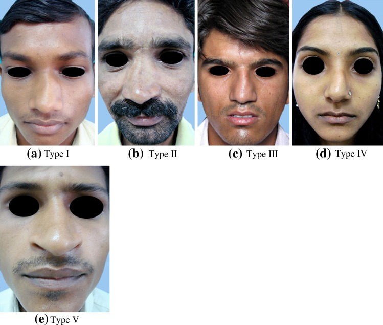
External nasal deformities were classified into I–V Types based on external deviation of horizontal subunits, the bony pyramid and cartilaginous vault as described by Jang et al. [4] (Fig. 2; Table 2). 67% had external deformity while 33 were found to have purely septal deviation. In those patients with external deformity, Type I in 26% was most commonly observed external deformity. Type II deformity was seen in 15% (Table 5). Figure 5a–e shows the prototype of each deformity.
Table 5.
Distribution of patients according to external nasal deformity
| Type of external nasal deformity | No. of patients | Percentage |
|---|---|---|
| No deformity | 33 | 33 |
| I- Straight tilted bony pyramid with tilted cartilaginous vault in the opposite direction | 26 | 26 |
| II- Straight tilted bony pyramid with concavely or convexly bent cartilaginous vault | 12 | 12 |
| III- Straight bony pyramid with tilted cartilaginous vault | 15 | 15 |
| IV- Straight bony pyramid with bent cartilaginous vault | 6 | 6 |
| V- Straight tilted bony pyramid and tilted cartilaginous dorsum in the same direction | 8 | 8 |
| Total | 100 | 100.00 |
Fig. 5.
External nasal deformity. a Type I. b Type II. c Type III. d Type IV. e Type V
By sequential correlation of the type of septal deviation associated with each type of external deformity it was found that Type I, III and V septal deviations were not associated with any external deformity. Type II septal deviation was seen more commonly with Type III external deformity. Type IV septal deviation was associated more frequently with Type I external deformity (Table 6).
Table 6.
Correlation of external nasal deformity with nasal septal deviation
| External deformity | Deviated nasal septum | Total | ||||||
|---|---|---|---|---|---|---|---|---|
| I | II | III | IV | V | VI | VII | ||
| No deformity | 5 | 10 | 5 | 0 | 7 | 0 | 6 | 33 |
| I | 0 | 1 | 0 | 12 | 0 | 6 | 7 | 26 |
| II | 0 | 1 | 0 | 3 | 0 | 3 | 5 | 12 |
| III | 0 | 7 | 0 | 2 | 0 | 1 | 5 | 15 |
| IV | 0 | 0 | 0 | 2 | 0 | 1 | 3 | 6 |
| V | 0 | 2 | 0 | 3 | 0 | 0 | 3 | 8 |
| Total | 5 | 21 | 5 | 22 | 7 | 11 | 29 | 100 |
χ2-value = 76.17, P-value < 0.0001, Significant
Though not the primary objective of our study, on evaluation of patients, we noted Concha Bullosa in 18%, accessory ostium in 8% and paradoxical middle turbinate in 6%. Another important finding of our study is 11% of our patients had chronic suppurative otitis media (8% unilateral and 3% bilateral).
Discussion
Many investigators have attempted to develop and evolve a classification for deviated nasal septum and associated external nasal deformities. A classification with etiological correlation has been suggested [5]. This classification simply divides nasal septum into anterior cartilaginous deviation and combined (cartilaginous and bony) septal deformity. The merit of this classification lies in the fact that, it helps to correlate the cause for nasal septal deflections. Anterior cartilage deviation is typically localized to the anterior quadrilateral cartilage and frequently associated with asymmetry of external bony pyramid and dislocation of cartilage off the anterior nasal spine. This deformity is common in new born delivered vaginally. Combined septal deformity involves all septal component including vomer bone, the perpendicular plate of ethmoid and quadrilateral cartilage. Deformities can include a spur at vomer ethmoid junction or a C- or S-shaped bending of cartilage. They are typically associated with the deformity of cheek, external nares, palate and malocclusion of teeth. Therefore combined nasal deformity is part of greater generalized facial deformity [5].
A more pragmatic classification has been evolved based on morphology, site and severity of deviations [6]. In yet another classification [7] septum has been divided into five areas i.e. attic, valvular, vestibular, turbinate and posterior choanal region. This division accurately describes the anatomical, operational and functional implication of septal deviations. Septal defects has further been classified in three types, simple, obstructive and impactive [7]. In a study which critically looked at 1,224 septal surgeries [8] suggested six different categories of septal deviations, from class A to F. All these classifications are too modest and basic to accurately describe the various septal deviation. Mladina [2] developed a system that divides septal deformity in seven types (Fig. 1; Table 1) which was modified by Janardhan et al. [3]. We employed this classification for septal deviations in our study.
External nasal deformities have also been classified by various investigators [9–11]. For the present study, we adopted a classification given by Jang et al. [4] which describes external nasal deformity in I–V Types (Fig. 2; Table 2).
The age predilection in the present study showed that majority of patients (34%) fell in the age group of 31–40 years followed by 29% in the 21–30 years with the mean age of 34.7 years. In various other studies, mean age of 31.5 years [4], 37 years [6], 33.5 years [8] has been reported. These findings by different investigators are in keeping with our findings In this study male to female ratio of 1.7:1 has been observed Ratio of 1.8:1 [6] and 2.2:1 [3] have been reported. Preponderance in males can be reasoned out by the fact that most common etiology of deviated nasal septum is nasal trauma which occur more frequently in males [12]. It was found that nasal obstruction is the most common symptom followed by nasal discharge. In one of the studies [8] nasal obstruction (71%) nasal discharge (41%), headache (20%) sneezing (15% and epistaxis (3%) were the symptoms in cases of DNS in order of their frequency.
Morphologically deviation of nasal septum in our study were stratified into seven types by adopting a classification described by Mladina [2] and modified by Janardhan et al. [3]. Type VII was the most common deviation in 29% followed by Type IV in 22%. In contrast to our findings, Janardhan et al. [3] found Type V (45%), as the most common septal deviation. Incidentally in our study Type V was least common i.e. in 7% only. We found caudal dislocation in 64%, buckling of septum in 51% and inferior turbinate hypertrophy in 55% of patients of deviated nasal septum.
The severity of deviation was graded into mild, moderate and severe categories based on the distance of deviated nasal septum from lateral nasal wall from convex side. Moderate deviation formed the major chunk (49%) followed by severe deviation (28%) and mild deviation (23%). Similar findings were recorded [6] wherein moderate deviation in 53% followed by mild (25%) and severe (22%) were reported.
In the present study, external nasal deformity was classified into I–V Types based on the external deviation of two horizontal subunits, the bony pyramid and cartilaginous vault [4] of the total 100 patients, 67% had external deformity (Fig. 5a–e). In one of the previous studies [13] 70% of cases of septal deviation coexisted with deformity of the external nose Concomitant septoplasties in 80% of their primary and secondary rhinoplasties were performed [14] implying coexistence of nasal septal deviation and external nasal deformity in as many cases.
Type I (Fig. 5a) external deformity was the most common (26%) followed by Type III in 15% in our study. In one of the previous studies [4], Type I deformity accounted for, maximum 24 patients (32%) of their series were noted thus endorsing our observation.
On correlating nasal septal deviation with external nasal deformity, we observed that Type I, III and V septal deviation were not associated with any external deformity. It is largely because deviations in this type are localized deviation and are not strong enough to pull the nasal dorsum to create external nasal deformity. Type II septal deviation were seen most commonly with Type III external deformity (Tilt of cartilaginous vault keeping the bony pyramid central, Fig. 5c). It can be cogently argued that Type II septal deviation is a vertical cartilaginous deviation that can pull the mid dorsum without tilting bony pyramid. Type IV septal deviation is an ‘S’ shaped anterior cartilaginous and posterior bony deviation on the opposite side. This was most commonly seen with Type I external deformity This is because ‘S’ shaped septal deviation pull the bony pyramid in the direction of posterior bony septal deviation and cartilaginous vault is pulled on the opposite direction along with anterior septal deviation. On applying Chi square test significant correlation is found between deviated nasal septum and external nasal septum (χ2-value = 76.17, P-value < 0.0001, Significant).
Thus the close association between external nasal deformity and nasal septal deviation points towards the fact that neither the septum nor the external deformity can be evaluated and treated separately if final outcome as regards nasal form and function is to be gratifying.
Conclusions
In our evaluative study of 100 patients of deviated nasal septum spanning over a period of nearly 2 years, majority of patients were in the age group of 31–40 years with the gender ratio of 1.7:1. Type VII was the most common type of deviation in 29%. In our series, 67% patients had external nasal deformity and of the 67% patients having external deformity, Type I deformity was most common (26%).
Another remarkable findings of our study being Type I, III, V septal deviations were not associated with external deviation Type II septal deviation were mostly associated with Type III external deformity (7%) and Type IV septal deviation were associated with Type 1 external deformity (12%).
We noted Concha Bullosa in 18%, accessory ostium in 8% and paradoxical middle turbinate in 6%. Noteworthy finding of our study is 11% of our patients had chronic suppurative otitis media (8% unilateral and 3% bilateral).
References
- 1.Beechius GJ. Nasal septoplasty. Otolaryngol Clin N Am. 1973;6:693–710. [PubMed] [Google Scholar]
- 2.Mladina R, Cujić E, Subarić M, et al. Nasal septal deformities in ear nose and throat patients: an international study. Am J Otol. 2008;29(2):75–82. doi: 10.1016/j.amjoto.2007.02.002. [DOI] [PubMed] [Google Scholar]
- 3.Janardhan Rao J, Vinaykumar EC, Ram Babu K, et al. Classification of nasal septal deviation—relation to sinonasal pathologies. Indian J Otolaryngol Head Neck surgery. 2005;57(3):199–201. doi: 10.1007/BF03008013. [DOI] [PMC free article] [PubMed] [Google Scholar]
- 4.Jang JY, Wang JH, Lee BJ. Classification of the deviated nose and its treatment. Arch Otolaryngol Head Neck Surg. 2008;134(3):311–315. doi: 10.1001/archoto.2007.46. [DOI] [PubMed] [Google Scholar]
- 5.Gray LP. Deviated nasal septum, incidence and etiology. Ann Otol Rhinol Laryngol. 1978;87(3 pt 3 supplement 50):3–20. doi: 10.1177/00034894780873s201. [DOI] [PubMed] [Google Scholar]
- 6.Jin HR, Lee JY, Jung WJ. New description method and classification system for septal deviation. J Rhinol. 2007;14(1):27–31. [Google Scholar]
- 7.Cottle MH. Structure and function of nasal vestibule. Arch Otolaryngol Head Neck Surg. 1955;62:173. doi: 10.1001/archotol.1955.03830020055011. [DOI] [PubMed] [Google Scholar]
- 8.Guyuron B, Uzzo C, Scull H. A practical classification of septonasal deviation and an effective guide to septal surgery. Plast Roconstr Surg. 1999;104:2202–2209. doi: 10.1097/00006534-199912000-00039. [DOI] [PubMed] [Google Scholar]
- 9.Brain D (1979) The nasal septum Scott Brown ‘s diseases of Ear Nose and Throat, vol 3. Butterworths and Co Ltd, London, pp 110–111
- 10.Ellis DA, Gilbert RW. Analysis and correction of crooked nose. J Otolaryngol. 1991;20(1):14–18. [PubMed] [Google Scholar]
- 11.Huizing EH, Groot JAM. Functional reconstructive nasal surgery. New York: Thieme’s NY book publication; 2003. pp. 55–59. [Google Scholar]
- 12.de Oliveira AKP, Elias Junior E, dos Santos LV, Bettega SG, Mocellin M (2005) Presence of deviated nasal septum in Curitiba, Brazil. Int Arch Otolaryngol 9(4):123
- 13.Goldman IB. New technique in surgeries of deviated nasal septum AMA. Arch Otolaryngol. 1956;64(3):183–189. doi: 10.1001/archotol.1956.03830150013003. [DOI] [PubMed] [Google Scholar]
- 14.Meyer R (2002) Secondary rhinoplasty including reconstruction of nose, 2nd edn. Springer, Berlin, pp 81


