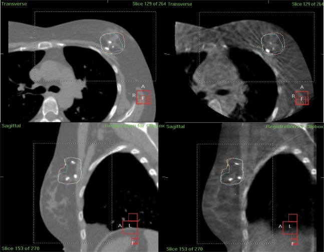
Figure 2.
An example of reference and verification images from a patient with left breast disease. Transverse view from reference CT scan (top left); transverse view from verification cone beam CT (CBCT) scan (top right); sagittal view from reference CT scan (bottom left); sagittal view from verification CBCT scan (bottom right). Images show tumour bed planning target volume (blue contour) and 95% isodose structure (pink structure).

