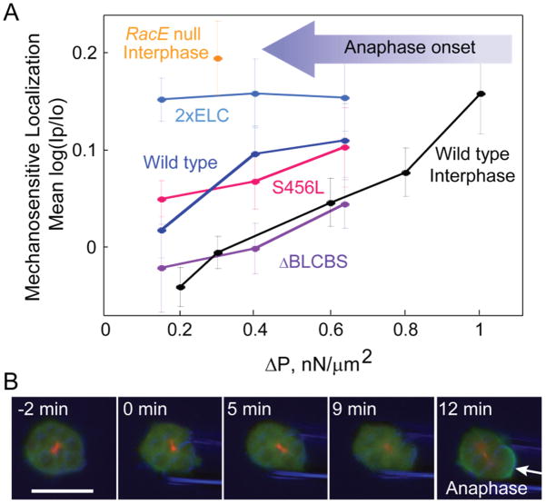
Figure 2. Myosin mechanosensation is dependent on the lever arm length and cell cycle phase (Effler et al. 2006; Luo et al. 2012; Ren et al. 2009).
A) Myosin localization to the pipette in dividing cells was measured under a range of aspiration pressures for various lever arm mutants, as well as for WT interphase cells. Upon anaphase onset (large arrow), the pressure regime (ΔP) required to trigger myosin II accumulation is shifted to the left. Mean log(Ip/Io) is the average log of the ratios of GFP-myosin II intensity (I) of the cortex inside the micropipette (Ip) to the opposite cortex (Io).
B) A continuously aspirated mitotic cell becomes mechanosensitive upon anaphase onset. The arrow identifies the mechanosensitive accumulation of myosin II. Green-GFP labeled myosin II, Red-RFP labeled tubulin, Blue-DIC.

