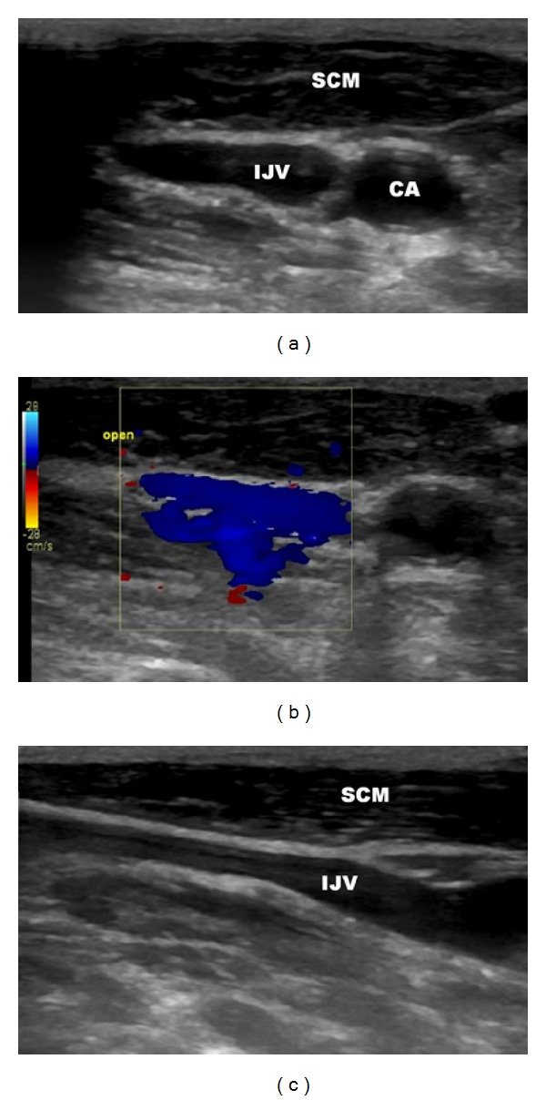
Figure 3.

Sonography of left the internal jugular vein before the surgical procedure, mouth opened. (a) transverse imaging, (b) transverse imaging, good flow through the vein as demonstrated by blue color, (c) longitudinal imaging; IJV: internal jugular vein, CA: carotid artery, SCM: sternocleidomastoid muscle.
