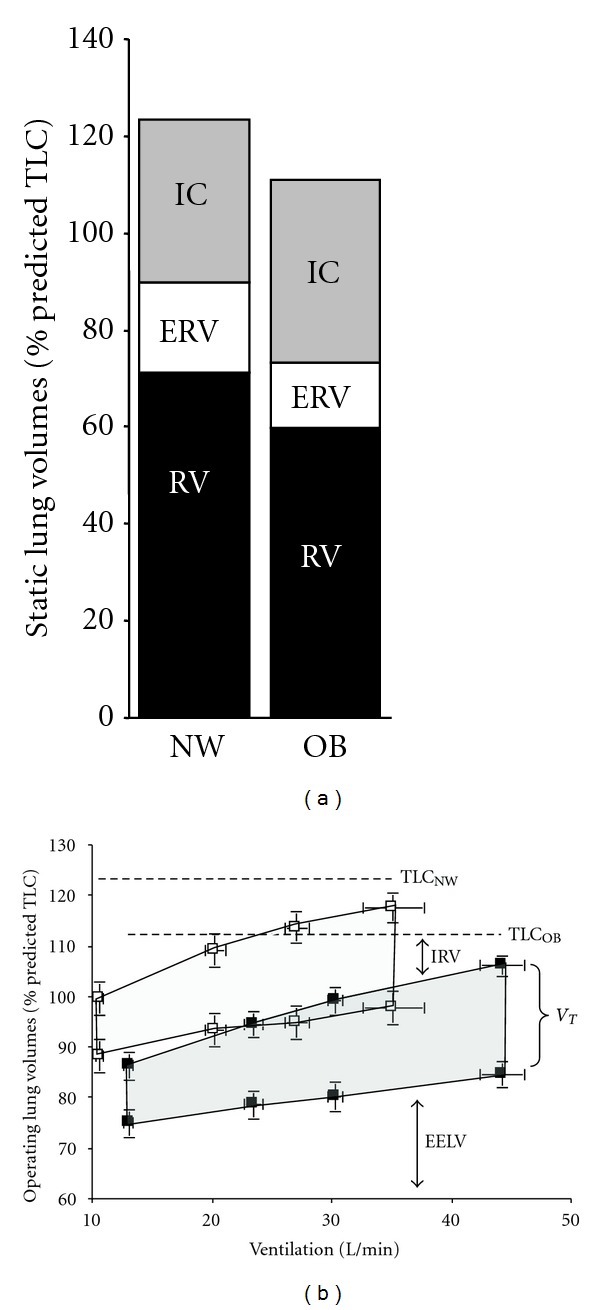
Figure 7.

(a) Static lung volumes measured by body plethysmography are shown at rest. Expiratory reserve volume (ERV) and functional residual capacity (FRC = ERV + RV) were significantly lower in the obese (OB) group compared with the normal weight (NW) group with COPD. (b) Operating lung volumes (mean ± SEM) are shown from rest-to-peak exercise in the OB (closed symbols) and NW (open symbols) subjects: end-expiratory lung volume (EELV) was consistently lower at rest and throughout exercise in OB; the OB group reached an EELV at peak exercise that was similar to that of the NW group at the preexercise resting level. IC: inspiratory capacity; IRV: inspiratory reserve volume; V T: tidal volume (shaded area); RV: residual volume, from Ora et al. [13].
