
Figure 3.
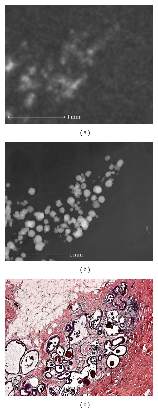
Fibrocystic mastopathy with sclerosing adenosis. Round microcalcification that is typically benign in microradiography (b) appears linear and amorphous in conventional specimen radiography as a result of the superposition (a). The calcification is marked by arrows in the histological picture (c).