
Figure 2.
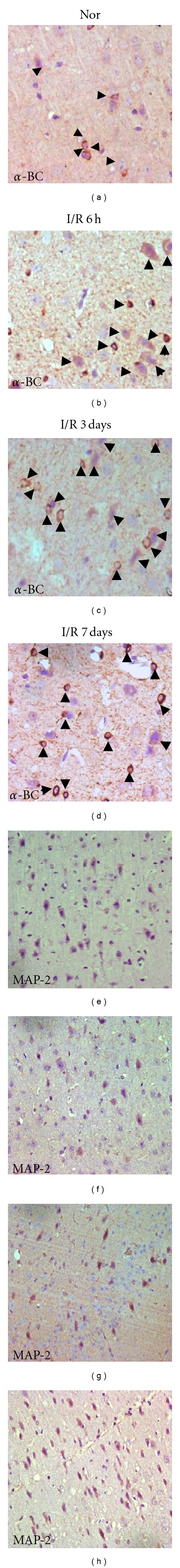
Immunohistochemical findings (DAB × 20) of α-BC or MAP-2 in Gerbil brain sections of normal (A/E) and ischemia/reperfusion (I/R) 6 hours (B/F), 3 days (C/G), and 7 days (D/H) after occlusion. An increment in the number of α-BC-positive cells (black arrow head) was detected 6 hours after reperfusion and lasted until the 7th day after reperfusion; an immediate reduction of MAP-2-positive neurons in 6 hours after reperfusion was detected and began to recover after 3 days post-reperfusion, on the 7th day the number of MAP-2-positive neurons was almost normal.