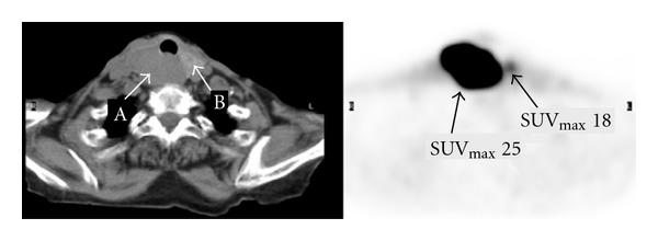
Figure 3.

Transaxial FDG PET-CT images of the neck show a large lesion with SUVmax 25 in the right lobe of the thyroid (arrow A). There is a 1.5 cm nodule with mild to moderate uptake in the left lobe, but unexpectedly measured SUVmax is 18 (arrow B).
