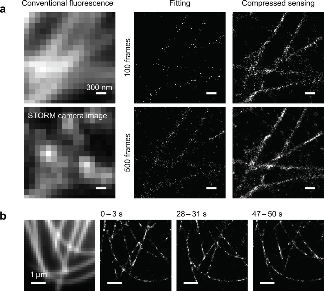
Figure 2.
Experimental STORM images using compressed sensing. (a) STORM imaging of microtubules in Drosophila S2 cells immunostained with secondary antibody labeled with the Alexa Fluor 647 - Cy3 dye pair. Left column: conventional fluorescence image and one raw image frame during STORM data acquisition, showing high density of activated fluorophores. Middle column: result of single-molecule fitting, reconstructed from 100 and 500 frames of camera images, respectively. Right column: result by compressed sensing using the same set of camera images. Scale bars: 300 nm. (b) STORM imaging of mEos2-tubulin in a living Drosophila S2 cell. The conventional fluorescence image in the leftmost panel is acquired before STORM imaging. Three snapshots from the STORM movie are displayed, each with 3 seconds integration time. The dynamics of the microtubules can be clearly observed. See Supplementary Video online.

