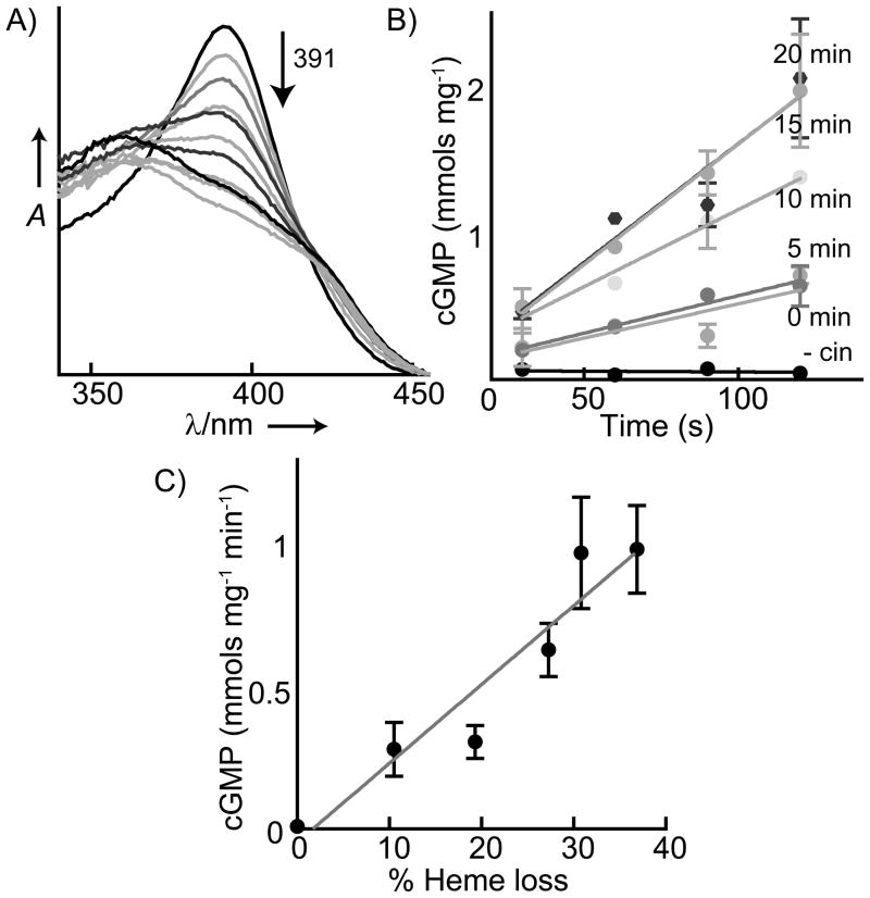
Figure 3.
Correlation of ferric sGC heme loss with activity in the presence of cinaciguat. A) Heme dissociation from ferric sGC (1.8 μM) in the presence of cinaciguat (20 μM) was observed via the decrease in the sGC Soret maximum (391 nm) with time. B) The initial rate of sGC activity is shown from aliquots withdrawn at the same time as spectra were taken in (A) (without cinaciguat: - cin). C) Correlation of the increase in ferric sGC activity with heme replaced by cinaciguat.

