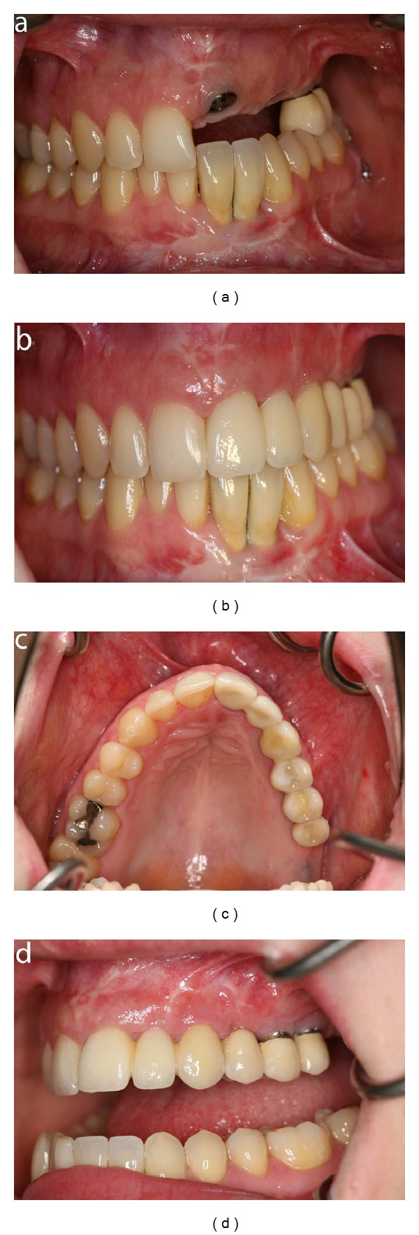
Figure 12.

Patient 2: intraoral photographs demonstrating the dentition during and after treatment with dental implants. (a) Frontal view before crowns are inserted on the implants in the left maxillary incisor, canine, and first premolar region. Crowns have been inserted in the left mandibular incisor region. (b) Frontal view after insertion of crowns on the implants. (c) Occlusal view after insertion of crowns on the implants. (d) Lateral view demonstrating the crowns inserted in the implants in the left side of the maxilla.
