Figure 3.
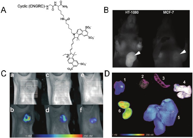
In vivo imaging with NGR-Cy 5.5. (A) Chemical structure of Cy 5.5-labeled NGR-peptide. (B) In vivo fluorescence reflectance imaging (FRI) 24 h after the injection of NGR-Cy 5.5 to HT-1080 and MCF-7 xenografts. Arrows indicated the tumors. (C) Top part: fluorescence-mediated tomography (FMT) 24 h after injection of NGR-Cy 5.5 to mice bearing (a) HT-1080, (e) MCF-7, (c) mice bearing HT-1080 that were pre-injected with100-fold unlabeled peptide 10 min ahead of injection with NGR-Cy5.5; Bottom part: FMT 60 min after injection of NGR-Cy5.5 to (b)(d)(f) that are prepared under the same conditions to (a)(c)(e), respectively. (D) Overlay of white light and FRI images for organs of HT-1080 bearing mice 24 h after injection of NGR-Cy5.5; (1) HT-1080 tumor (2) heart (3) spleen (4) lung (5) liver (6) kidneys. Adapted from reference [18].

