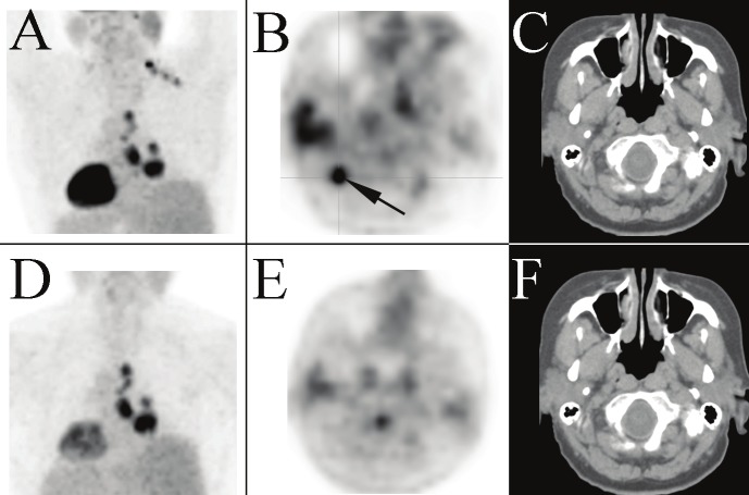
Figure 2.
Posterior upper body view of the MIP from a PET/CT performed on a 61 year old woman (patient 5) to stage a suspected primary lung cancer (A). A unilateral cluster of distinct foci are noted at the base of the neck extending down to the supraclavicular region on the right side. The most intense supraclavicular focus is shown on the transaxial PET image (B). There is no corresponding lymph node on the corresponding transaxial CT image (C), presumably hypermetabolic BAT. The right mediastinal activity on the MIP is consistent with the patient’s known disease. The BAT activity resolves on the post-diazepam PET/CT study repeated three days later; the MIP (D) and transaxial PET (E) and CT (F) images of the neck are provided.

