
Figure 4.
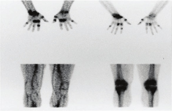
Images obtained 4 h after injection of 99mTc-1.2B6-Fab (left) and 99mTc-HDP (right) in two patients with RA. The images on the top correlate well; the bottom images show discordance between the lack of uptake of the mAb fragment and diffuse bony uptake. (Reprinted from Jamar F, Houssiau FA, Devogelaer JP, Chapman PT, Haskard DO, Beaujean V, Beckers C, Manicourt DH and Peters AM. Scintigraphy using a technetium-99m labelled anti-E-selectin Fab fragment in rheumatoid arthritis. Rheumatology (Oxford) 2002; 41: 53-61; by permission of Oxford University Press).