
Figure 1. Diagram of the model.
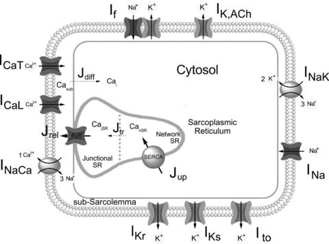
Schematic diagram of the model depicting cell compartments and major functional components. Fluid compartments include cytosol, subsarcolemmal space and sarcoplasmic reticulum (network and junctional SR). Membrane currents are shown with associated ion selectivities (see Supplemental Material for abbreviations). The Ca2+-handling system comprises Ca2+ diffusion from submembrane space to myoplasm (Jdiff), Ca2+ uptake by SR (Jup), Ca2+ pump (SERCA), transfer between network and junctional SR (Jtr), and release (Jrel) by ryanodine receptors (RyRs).