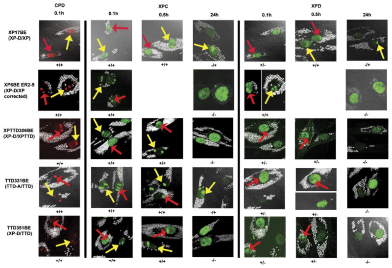
FIGURE 3.
Recruitment of XPC and XPD proteins to localized DNA damage in XP, XP/TTD, and TTD cells at 0.1 hr, 0.5 hr, and 24 hr after UV irradiation. Normal cells (AG13145) were labeled with 0.8-μm latex beads (red arrows) and cells (from Patients XP17BE, XP6BE ER2-9, XPTTD306BE, TTD331BE, and TTD351BE) were labeled with 2-μm latex beads (yellow arrows). UV irradiation at 100 J/m2 was delivered through a 5-μm filter. Cells were fixed by 0.1 hr, 0.5 hr, or 24 hr postirradiation and immunostained with pairs of antibodies to simultaneously assess the location of DNA damage and NER proteins. The arrows indicate sites of localized immunostaining: normal (red arrows), patient (yellow arrows). Where necessary, images were captured at two exposure times to show any localization with low levels of protein (images separated by white line). Symbols below each image indicate localization (+) or nonlocalization (−) of NER proteins or photoproducts in normal cells/patient cells.

