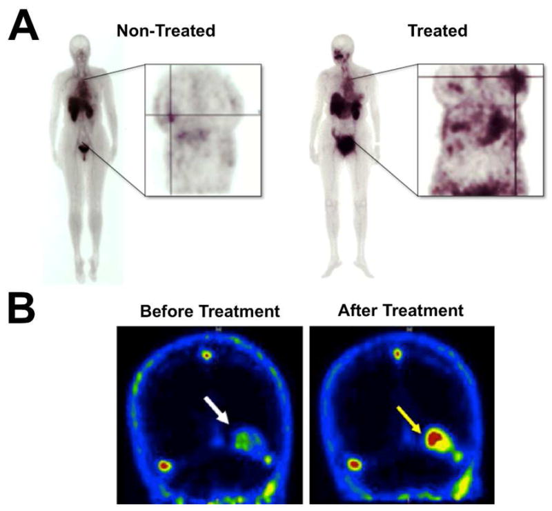
Figure 5.

Clinical imaging with cell death molecular probes. (A) Scintigraphic images of separate non-treated and drug treated breast cancer patients who were injected with 99mTc-EC-Annexin V. Annexin V had higher accumulation in treated breast tumors compared to non-treated tumors, though the probe localized to both tumors as indicated by the cross-lines. (B) 18F-ML-10 PET images of a brain metastasis in a single patient (indicated by arrow) before and after whole brain radiation therapy. The low level of 18F-ML-10 accumulation in the metastasis prior to treatment likely represents a basal amount of cell death. Reprinted with permission from references 89 and 147.
