
Fig. 99.
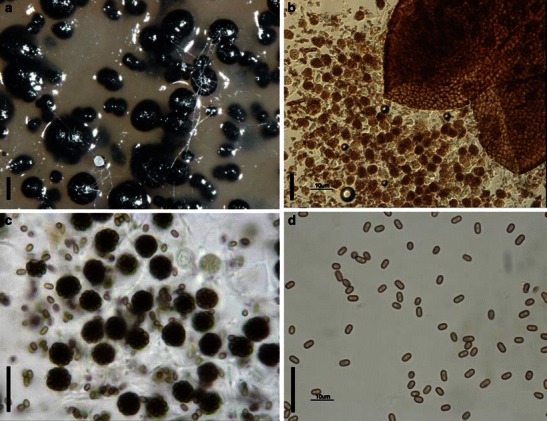
Pycnidiophora dispersa (A from CBS 297.56; B-D from MSC 133.118, type). a Ascomata scattering on the surface of the substrate. b Crashed ascoma. Note the numerous released asci. c Globose asci and released ascospores. d One-celled ascospores. Scale bars: a = 200 μm, b–d = 20 μm